Abstract submission for the 17th Annual Meeting of the Organization for Human Brain Mapping to be held in Québec City, Canada, June 26-30, 2011 is now open. The deadline to submit has been extended to January 10, 2011. Please note that this deadline is firm, there will be no further extensions.

Wednesday, December 29, 2010
some updates
I located the bug with some ROI's TFR permutation test (TFR data not-assigned error only for induced response, but I don't think I have time to fix it right now. I am work on the full spectrum data for the current work, if it is necessary to look at the induced response, then I will have to fix the bug myself).
Filling in this table -- I'm working on the data full time tomorrow, should be able to finish this and assemble the figs by mid night.
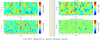
Left ACC activation:
Writing some scripts to plot individual TFR for left ACC
close look at 1-40 Hz:
Right ACC
hi-gamma started from around 1 s after A onset
L_SMC
Sunday, December 26, 2010
Re: MEGANTI notes
I think it should be a good idea to use the same protocols for naming, folder structures, and especially important for the naming of Matlab scripts if we analyze different MEG projects under one server. This fig shows the steps for analysis, will show more detail when I have time.
MEG dataset are under folder:
/root/MEGANTI/data/XX
XX is each subject's initial.
Preprocessing first step: Marker files.
It is to find out the events of interest and save the timestamps of each event into text files for further epoching the datasets (for example, "Instruction", "GO_cue", etc.
Each subject's main marker file "MarkerFile.mrk" is under each subject's raw MEG dataset. e.g. subject AB's main marker file is under:
/root/MEGANTI/data/AB/S07_MEG54_VEF_02.ds/
The marker file could be open with any text editor.Please refer to Jason's notes for the meaning of the events in the MarkerFile.mrk ("LightOn", "LightOff"...) and Presentation's log files.
I usually copy each subject's main marker file to the folder where I save Presentation's log file, e.g. /root/MEGANTI/data/00_gain/XX (XX is subject's initial, I think I only did for subject SW). After I am done with all the event's marker, I will copy the marker files (e.g. Instruction.txt, Go_cue.txt) to /root/MEGANTI/data/XX/markers
You only need two columns in each event's marker file, first column is the trial number, the second column is the offset from where the current trial begins (this "trial" is the trial length for MEG recording = 20 s/trial, not the actual the experiment's trial length).
e.g.
Marker file "Instruction.txt" should be something like this:
+0 +4.273600
+2 +3.158400
+2 +13.193600
+2 +17.808000
+3 +2.544000
+5 +2.974400
+6 +7.227200
+6 +16.692800
+7 +0.710400
+9 +2.710400
+9 +11.494400
+11 +12.478400
+15 +4.347200
+16 +19.947200
+17 +13.998400
+18 +3.448000
+19 +9.979200
Second step: adding markers and epoching
All the linux scripts are saved under:
/root/MEGANTI/data/02_scripts/
After we are done with all the event's marker files for each subject, we should then copy each subject's marker files (e.g. Instruction.txt, Go_cue.txt) to /root/MEGANTI/data/XX/markers. Next, we will need to write scripts for adding the markers to the raw dataset and epoching the data.
(a) Adding the markers:
e.g. For subjecg SW, we first log onto the Linux console with root account, then run:
export dataHome=~/MEGANTI/data
cd $dataHome/SW
export RAW_DS=S02_MEG54_VEF_02.ds
addMarker -f -n Instructions -l red -p markers/Instructions.txt $RAW_DS
(This marker "Instructions" would now be added to the main marker file)
(b) Epoching. After adding the markers, we run newDs to generate new datasets:
newDs -f -overlap 3 -marker "Instructions" -time -2 2.5 $RAW_DS SW_Instructions_-2.25_2.45.ds
Third step: Creating multi-sphere head models
To be continued...
Fiducial markers are under:
/root/MEGANTI/doc/fiducial/
Note:
(1) The MRI files need to be converted into CTF v2 format .mri file before we could run the headmodeling scripts
(2) The head modeling scripts must be run from Matlab GUI.
(3) Convention: the .mri file should be under each subject's home folder (/root/MEGANTI/data/XX/), the raw dataset usually are taken as the default dataset for creating head models.
e.g. makeHeadModels('/root/MEGANTI/data/SW/SW_V2.mri', '/root/MEGANTI/data/SW/S02_MEG54_VEF_02.ds')
Fourth step:run event-related beamformers:
To be continued...
MEG dataset are under folder:
/root/MEGANTI/data/XX
XX is each subject's initial.
Preprocessing first step: Marker files.
It is to find out the events of interest and save the timestamps of each event into text files for further epoching the datasets (for example, "Instruction", "GO_cue", etc.
Each subject's main marker file "MarkerFile.mrk" is under each subject's raw MEG dataset. e.g. subject AB's main marker file is under:
/root/MEGANTI/data/AB/S07_MEG54_VEF_02.ds/
The marker file could be open with any text editor.Please refer to Jason's notes for the meaning of the events in the MarkerFile.mrk ("LightOn", "LightOff"...) and Presentation's log files.
I usually copy each subject's main marker file to the folder where I save Presentation's log file, e.g. /root/MEGANTI/data/00_gain/XX (XX is subject's initial, I think I only did for subject SW). After I am done with all the event's marker, I will copy the marker files (e.g. Instruction.txt, Go_cue.txt) to /root/MEGANTI/data/XX/markers
You only need two columns in each event's marker file, first column is the trial number, the second column is the offset from where the current trial begins (this "trial" is the trial length for MEG recording = 20 s/trial, not the actual the experiment's trial length).
e.g.
Marker file "Instruction.txt" should be something like this:
+0 +4.273600
+2 +3.158400
+2 +13.193600
+2 +17.808000
+3 +2.544000
+5 +2.974400
+6 +7.227200
+6 +16.692800
+7 +0.710400
+9 +2.710400
+9 +11.494400
+11 +12.478400
+15 +4.347200
+16 +19.947200
+17 +13.998400
+18 +3.448000
+19 +9.979200
...
Second step: adding markers and epoching
All the linux scripts are saved under:
/root/MEGANTI/data/02_scripts/
After we are done with all the event's marker files for each subject, we should then copy each subject's marker files (e.g. Instruction.txt, Go_cue.txt) to /root/MEGANTI/data/XX/markers. Next, we will need to write scripts for adding the markers to the raw dataset and epoching the data.
(a) Adding the markers:
e.g. For subjecg SW, we first log onto the Linux console with root account, then run:
export dataHome=~/MEGANTI/data
cd $dataHome/SW
export RAW_DS=S02_MEG54_VEF_02.ds
addMarker -f -n Instructions -l red -p markers/Instructions.txt $RAW_DS
(This marker "Instructions" would now be added to the main marker file)
(b) Epoching. After adding the markers, we run newDs to generate new datasets:
newDs -f -overlap 3 -marker "Instructions" -time -2 2.5 $RAW_DS SW_Instructions_-2.25_2.45.ds
Third step: Creating multi-sphere head models
To be continued...
Fiducial markers are under:
/root/MEGANTI/doc/fiducial/
Note:
(1) The MRI files need to be converted into CTF v2 format .mri file before we could run the headmodeling scripts
(2) The head modeling scripts must be run from Matlab GUI.
(3) Convention: the .mri file should be under each subject's home folder (/root/MEGANTI/data/XX/), the raw dataset usually are taken as the default dataset for creating head models.
e.g. makeHeadModels('/root/MEGANTI/data/SW/SW_V2.mri', '/root/MEGANTI/data/SW/S02_MEG54_VEF_02.ds')
Fourth step:run event-related beamformers:
To be continued...
re: matlab scripts notes
permute_vs_tfr_sheng.m logic:
modified its code and export all the data to mat files.
new function for plotting permute_tfr:
function plot_permutation_tfr_sheng(perm_matFile, freqWindow, timeWindow )
need to find bugs in getting raw files (e.g. B002 L_ST_MT_BA39)
modified its code and export all the data to mat files.
new function for plotting permute_tfr:
function plot_permutation_tfr_sheng(perm_matFile, freqWindow, timeWindow )
need to find bugs in getting raw files (e.g. B002 L_ST_MT_BA39)
Friday, December 17, 2010
Visual alpha -- to be continued
G.Pfurtscheller 1994 paper gives a good explanation event-related desychronization (ERD) in alpha band in the visual system. (Pfurtscheller is the person coiled term ERD)
"Two different types of ERD can be differentiated: one short-lasting, localized to occipital areas and involving upper alpha components; the other longer lasting, more widespread, most prominent over parietal areas and maximal for lower alpha components. The former most likely reflects primary visual processing and feature extraction, the latter is more related to cognitive processing and mechanisms of attention. "
N. Yamagishi (2005) reported attentional shifts modifies the latency and the amplitude in MEG induced responses in cuneus region (BA17).
Pfurtscheller, G., C. Neuper, et al. (1994). "Event-related desynchronization (ERD) during visual processing." International Journal of Psychophysiology 16(2-3): 147-153.
Yamagishi, N., N. Goda, et al. (2005). "Attentional shifts towards an expected visual target alter the level of alpha-band oscillatory activity in the human calcarine cortex." Cognitive Brain Research 25(3): 799-809.
"Two different types of ERD can be differentiated: one short-lasting, localized to occipital areas and involving upper alpha components; the other longer lasting, more widespread, most prominent over parietal areas and maximal for lower alpha components. The former most likely reflects primary visual processing and feature extraction, the latter is more related to cognitive processing and mechanisms of attention. "
N. Yamagishi (2005) reported attentional shifts modifies the latency and the amplitude in MEG induced responses in cuneus region (BA17).
Fig. 4. Group analysis (n = 14) showing event-related spectral perturbation (ERSP) plots derived from the activation waveforms of the ICs reflecting calcarine activity for each subject and for each experimental condition (ISI = 1000 ms). The plots consist of 25 equally spaced bins with center frequencies ranging from 1.95 to 48.8 Hz and 32 equally spaced time steps with centers at 72 to 1372 ms. The group-mean ERSP plots show postcue power differences (in dB) referenced to a 200 ms precue baseline recording for (a) attention directed away from the stimulus and (b) attention directed towards the stimulus. Both spectral power increases (reddish hues) and decreases (bluish hues) are evident. A random effects one-sample t test indicating significant ( P < 0.05 FDR corrected; df = 13) positive (reddish hues) and negative (bluish hues) event-related spectral power changes are shown for (c) attention directed away from the stimulus and (d) attention directed towards the stimulus. The cue onset time (t = 0 ms) and the stimulus onset time (t = 1000 ms) are indicated by vertical dotted lines in each plot.
==> different in virtual sensor power could imply attention directed to the stimulus (or the subjects were more vigilant in the correct trials), and thus more likely to perceive the change in speed -- a reasonable explanation? The following is an old fig that aligned to the onset of stimulus in left VF display, the high alpha (around 8 - 12 Hz) shows power difference when stimulus is on. I modified the scripts to get the data in mat format for and will soon modify the figs as soon as the data are generated.
Pfurtscheller, G., C. Neuper, et al. (1994). "Event-related desynchronization (ERD) during visual processing." International Journal of Psychophysiology 16(2-3): 147-153.
Yamagishi, N., N. Goda, et al. (2005). "Attentional shifts towards an expected visual target alter the level of alpha-band oscillatory activity in the human calcarine cortex." Cognitive Brain Research 25(3): 799-809.
Wednesday, December 15, 2010
Right ACC following B (speed change)- Change P = .01
R ACC (13 12 45) mag. = 0.71 @230 ms

Look up
What is the lower resolutaion in time of the low freq domain... (previous figure)
------------------------
What is happening at the medial activation 60 ms - what is at medial/cingulate activation?
V. van Veen & Carter 2002: following impulsive errors (response made why stimulus evaluation is imcomplete), a large negative deflection (ERN, the detection that an error was made)
==> We did not exclude the correct trials that might be impulsive responses (small response latency)
Caudal ACC:
* frontalcentral N2 prior to the response on correct trials with stimulus conflict, 340 - 380 ms after onset. (response latency 450 - 500 ms)
* ERN immediately following error response (40 - 80 ms)
Rostral area of ACC:
* error related positivity peaking at 200 -250 ms following ERN
ACC is also strongly activated during "guilty knowledge" & "lies"
Actually the medial frontal/ACC activation magnitude at 50 ms is higher (reported as superior frontal gyrus, more medial 2 voxel superior to the peak found at 60 ms, spatial resolution 2.5 mm):
@50 ms (-10 11 55) mag = 0.73
@60 ms (-18 10 50) mag = 0.68
HBM - do methods section...
Write abstract - than make figures that prove the story.
Friday, December 10, 2010
Wednesday, December 8, 2010
Movies avg for both left and right hemifield display
B002: speed change onset @ 0 ms, the change last 200 ms
A002: GO cue @ 2200 ms, Stimulus onset @ 0 ms
C002: Key pressing
Thursday, December 2, 2010
Comparison of correct and error activations
Will try to get individual glassbrain, and search for early premotor activation
After B (speed change)
After A002 (GO cue)
Early motor premotor response after speed change
Have stats class tomorrow, but will look at the permute_tfr program follow Paul's hint in the afternoon. Try to fix the error in the code.
50 ms after speed change in correct trials, superior temporal activation, then @ 120 ms motor & premotor, parallel network the activation of motor response even before the change was perceived? Need to look at individual data for this effect?
50 ms after speed change in correct trials, superior temporal activation, then @ 120 ms motor & premotor, parallel network the activation of motor response even before the change was perceived? Need to look at individual data for this effect?
@ 210 ms
@ 220 ms
@ 250 ms
@ 280 ms
Left SMC peak picked from 120 ms after B onset. There is signal in Left SMC after the change onset, but power increase in error around key pressing are large -- can't get the induced TFR due to error in TFR plotting code.
Wednesday, December 1, 2010
Sunday, November 28, 2010
[B002] Permutation: starting from MT+? @220 ms ==> right IPL @250 ==> left IPL @ 280
Aligned to Event B
(speed change onset)
The permutation test for the glass brain should be done by Monday noon for the event of B, A002 (GO cue) and C (key down), then the permutation threshold for each time slice will be used to search activation peaks in Tal. space for all the three events.
Correct Trials
@220 ms activation at left MT+
250 ms peak reported at right IPL
@280 ms, left IPL
Error trials
at the same time slice
@220 ms
@230 ms
@250 ms
@280 ms
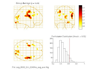
[A002] Permutation: 40 ms after "Go" cue, parietal and frontal activation
All the averaged figs shown here are based on both left and right visual field display. The label at the bottom of the fig indicates the event of interest (e.g. A002), the condition(correct or error), the time slice(2240 ms)
Note: A002 event (GO cue, the stimulus offset) always happens 2200 ms after the stimulus onset.

error trials: IPL activation moves around
Right premotor activated again @ 170 ms after GO, but correct trials has precuneus (KO?) activation.
Friday, November 26, 2010
Subscribe to:
Posts (Atom)


